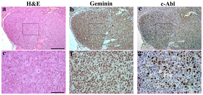
Figure 9. Histological and immunohistochemical analysis of geminin overexpressing mammary tumors.
(a and e) Representative H&E stained sections from induced Gem9 orthotopic mammary tumors. (b and C) and (f and g) adjacent sections to those shown in (a and e) stained with geminin (b and f) or c-Abl (c and g). Scale bars in a-c = 500 µm and d-f = 100 µm.

