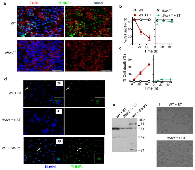
Figure 2.
Ifnar1−/− macrophages are resistant to S. Typhimurium–induced cell death. (a) Confocal microscopy of frozen sections of spleens obtained from wild-type and Ifnar1−/− mice 5 d after intravenous infection with 1 × 10² S. Typhimurium and stained for F4/80 and by TUNEL. Scale bars, 20 μm. (b,c) Viability, assessed by uptake of neutral red (b), and death, assessed by lactate dehydrogenase–release assay (c), of wild-type and Ifnar1−/− bone marrow–derived macrophages plated in 96-well plates at a density of 1 × 10⁵ cells per well and infected with S. Typhimurium (MOI, 10), then treated with gentamicin at 30 min after infection and assessed at 6, 24 and 48 h after infection. (d) Fluorescence microscopy of bone marrow–derived macrophages infected for 24 h with S. Typhimurium (+ ST) or treated for 3 h with staurosporine (+ stauro), then stained by TUNEL (green) and Hoechst nuclear dye (blue). Inset, enlargement of areas indicated by arrows. Original magnification, ×20 (main images) or ×60 (insets). Numbers in images indicate percent TUNEL+ cells (n ≥ 100 cells). (e) Immunoblot analysis of macrophages infected for 24h with S. Typhimurium or treated for 3 h with staurosporine, probed with antibody to cleaved PARP-1. (f) Fluorescence microscopy of bone marrow–derived macrophages infected for 24h with S. Typhimurium and stained with propidium iodide. Original magnification, ×20. Data are representative of three experiments with similar results (mean and s.e.m (b,c)).

