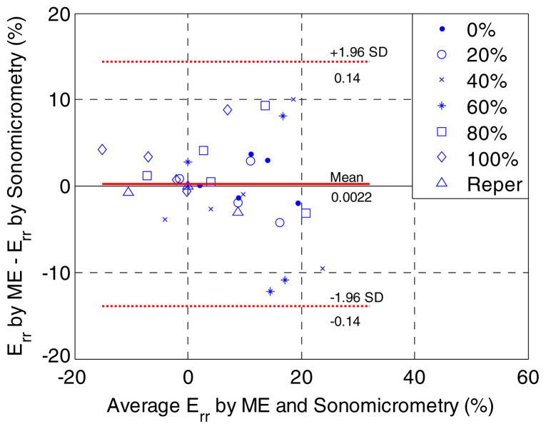
Figure 7.
A Bland–Altman plot of the end-systolic radial strains (Err) of five canine left ventricles in an anterior wall region of 3 × 3 mm2 at six different LAD flow reduction levels and reperfusion (Reper). SD denotes standard deviation. The bias and 95% limits of agreement were found to be 0.22% strain and −13.9% to 14.3% strain, respectively.

