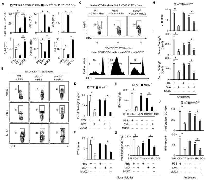
Fig. 3. MUC2 enhances gut homeostasis and oral tolerance.
(A) FC of CD103 and qRT-PCR of Il12a, Il12b, Il10, Aldh1a1 and Tgfb1 in SI-LP CD103+ DCs from WT or Muc2−/− mice gavaged for 5 days with phosphate buffer solution (PBS) or MUC2. RE, relative expression compared to Gapdh. (B) FC of Foxp3, IFN-γ, IL-17 and CD4 in SI-LP T cells from WT or Muc2−/− mice treated as in (A). (C) FC of Foxp3 and CD4 in naïve OT-II cells cultured for 5 days with SI-LP CD103+ DCs from WT or Muc2−/− mice treated as in (A) and intragastrically immunized with OVA. CD4+CD25+ OT-II cells from these cultures were incubated for 5 days with CFSE-labeled naïve OT-II cells and Abs to CD3 and CD28; divided CFSElow cells were quantified by FC. (D) ELISA of fecal OVA-specific IgG from WT and Muc2−/− mice tolerized with PBS, OVA or OVA plus MUC2 for 5 days and immunized as in (C). (E) ELISA of IFN-γ from OT-II cells incubated for 5 days with MLN CD103+ DCs from WT or Muc2−/− mice tolerized and immunized as in (D). (F) OVA-induced DTH in WT or Muc2−/− mice tolerized as in (C) and subcutaneously immunized with OVA. (G) ELISA of proliferation-induced BrdU from SPL CD4+ T cells activated for 5 days with OVA-pulsed SPL DCs from WT or Muc2−/− mice tolerized and immunized as in (F). (H–J) DTH, OVA-specific serum IgG and IgE, and SPL CD4+ T cell proliferation and IFN-γ secretion in WT or Muc2−/− mice immunized and tolerized as in (F) after oral antibiotics. Data summarize 2 experiments with ≥4 mice/group (error bars, s.d.; unpaired Student’s t test, *P <0.05) or show one of 4 experiments with similar results.

