
Figure 1.
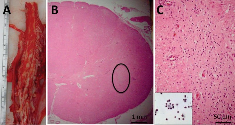
Neuropathological findings in Subject 14. (A) Gross image of cervical spinal cord at the time of autopsy. Serial sections through the region of transplantation did not demonstrate regions of cystic change, hemorrhage, or significant tissue disruption. (B) Representative cross section showing intact cord morphology using hematoxylin and eosin (H&E) staining. There is a nest of cells (circled) that are not intrinsic to the spinal cord, and do not stain with glial or neuronal markers (not shown). (C) Higher power of circled region in B showing the morphology of these cells, which is reminiscent of the morphology of the stem cells prior to transplantation (inset, H&E).