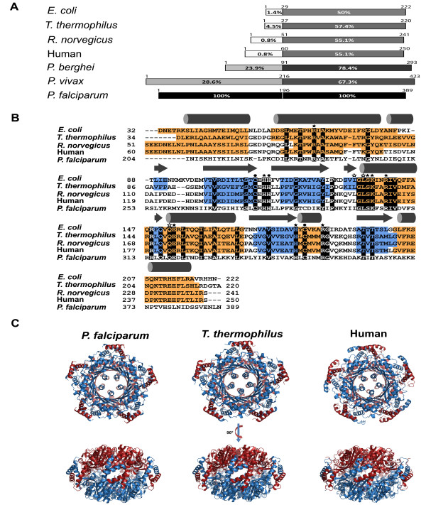
Figure 2.
Comparison of the GCH1 protein from Plasmodium falciparum to GCH1 proteins with known structures. (A) Sequence alignment of the GCH1 proteins. The GCH1 sequences were divided into the N-terminal regulatory domain and the C-terminal enzymatic core with the residue numbers on each diagram. The homology scores compared to P. falciparum GCH1were shown as percent homology and colour shade (100% and black colour to its own sequence). (B) Secondary structure diagram from known GCH1 structures and the homology model of P. falciparum GCH1 with α-helices in orange and β-strands in blue. The secondary structure diagrams of the P. falciparum GCH1 model are at the top of the alignment. The conserved amino acid residues are highlighted in black with labelled key residues (see text for detail). (C) Comparison of the overall homodecameric GCH1 structures. Two face-to-face pentameric rings are coloured in red and blue.

