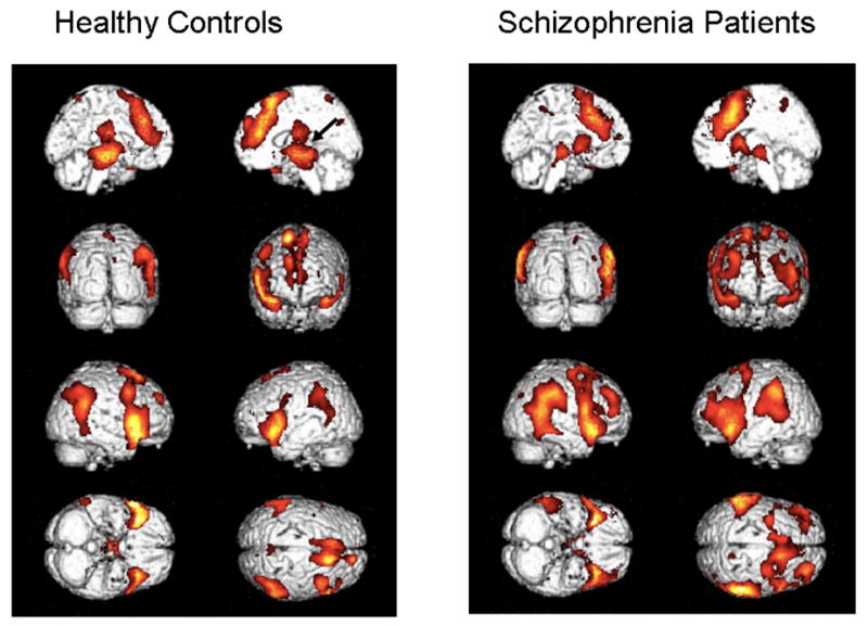
Figure 4.

Contrasts (p<.05, uncorrected) projected onto rendered brains to enable better identification of lateral activity. Healthy controls appear on the left and patients on the right. The left and right mid-sagittal views are seen in the 1st row, backward and forward facing views are seen in the 2nd row, right and left sides are seen in the 3rd row, and bottom and top views are seen in the 4th row.
