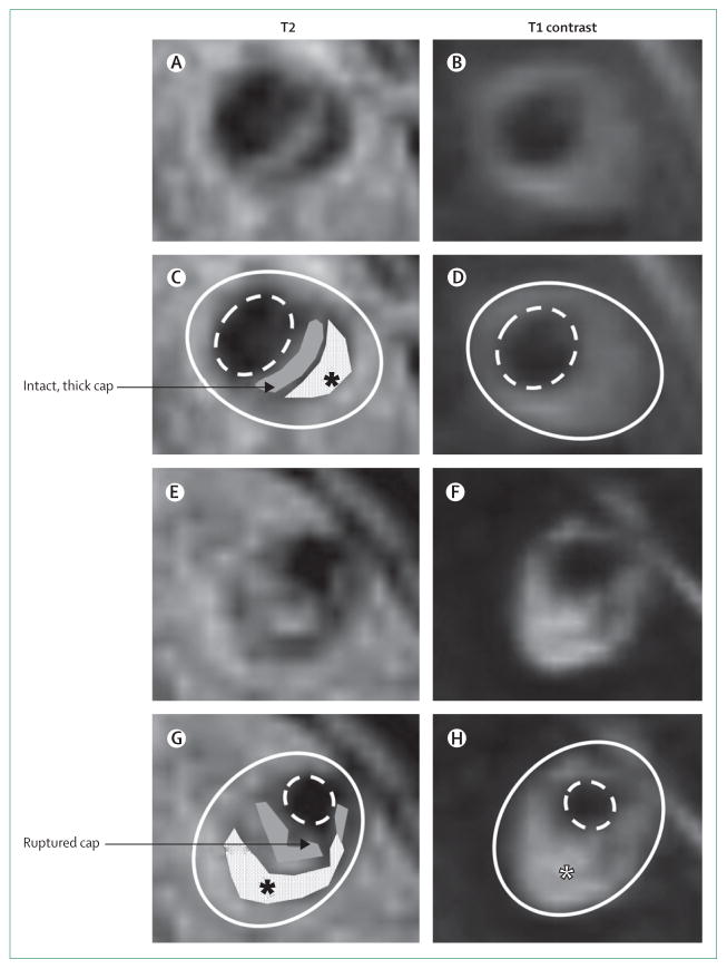
Figure 4. High-resolution MRI of vertebral artery stenoses with plaque components.
Panels A–D show T2-weighted and T1 post-contrast images (panels C and D have plaque components marked) of a cross-section of a vertebral artery plaque with a thick, intact, fibrous cap (grey) and lipid core (white with black asterisk). Panels E–H show T2-weighted and T1 post-contrast images (panels G and H have plaque components marked) of a cross-section of a vertebral artery plaque with a ruptured fibrous cap (grey) and lipid core (white with black asterisk), which enhances with contrast (white asterisk) and is also indicative of plaque rupture. The solid white line shows the outside vessel wall and the dashed white line the lumen.

