
Figure 2.
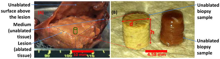
(A) Image of a 15×15 mm2 inclusion composed of a group of thermally-ablated lesions obtained by raster HIFU ablation on canine liver tissue ex vivo, (B) Representative biopsy samples from unablated and ablated canine liver tissues.
Official websites use .gov
A
.gov website belongs to an official
government organization in the United States.
Secure .gov websites use HTTPS
A lock (
) or https:// means you've safely
connected to the .gov website. Share sensitive
information only on official, secure websites.
(A) Image of a 15×15 mm2 inclusion composed of a group of thermally-ablated lesions obtained by raster HIFU ablation on canine liver tissue ex vivo, (B) Representative biopsy samples from unablated and ablated canine liver tissues.