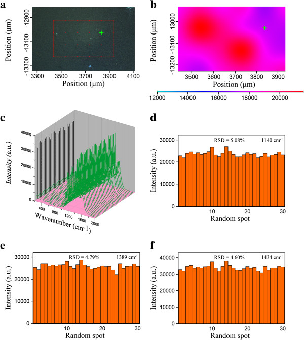
Figure 6.
Optical image, SERS mapping, SERS spectra, and RSD values. (a) Optical image of an area of 0.5 mm × 0.3 mm for the Ag/rGO nanocomposite 8C substrate. (b) The corresponding two-dimensional SERS mapping after 4-ATP adsorption. The peak mapped was at 1,140 cm−1. (c) A series of SERS spectra randomly collected from 30 spots of the Ag/rGO nanocomposite 8C substrate at 10−5 M 4-ATP. (d to f) The intensities of three main vibrations at 1,140, 1,389, and 1,434 cm−1 in the SERS spectra as shown in (c).

