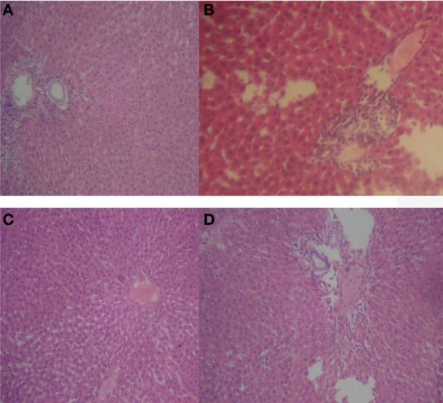
Figure 5.

Representative sections of rat liver in respect of reversibility study. (A) Normal rat liver showing normal hepatocytes and hepatic vasculature; (B) 20 mg/kg/day HLE-FP-treated rat liver showing epithelial proliferation and scattered areas of vascular congestion; (C) 100 mg/kg/day HLE-FP-treated rat liver showing scattered areas of vascular congestion; and (D) 500 mg/kg/day HLE-FP-treated rat liver showing few local lymphocytic aggregates with mild-to-moderate vascular congestion (× 100 magnification).
