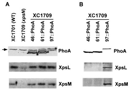
FIG. 3.
(A) Immunoblot analysis of XpsN::PhoA fusion proteins. Total cell lysates of X. campestris pv. campestris were analyzed by separating them on SDS-PAGE, followed by Western blotting using anti-PhoA antibody (top), anti-XpsL antibody (middle), or anti-XpsM antibody (bottom). Each XpsN::PhoA fusion protein is designated by the position, in amino acid residues, at which PhoA is fused. The asterisks mark the protein bands of the fusion proteins. The extra protein band (indicated by an arrow) appearing in all samples is probably a cross-reactive material detected by the anti-PhoA antibody. (B) Coimmunoprecipitation of XpsN::PhoA fusion proteins with XpsL and XpsM. Membrane vesicles prepared from XC1709 that expresses different XpsN::PhoA proteins were extracted with 2% Triton X-100, followed by precipitation with anti-PhoA antibody. The immunoblots were detected with anti-PhoA antibody (top), anti-XpsL antibody (middle), and anti-XpsM antibody (bottom).

