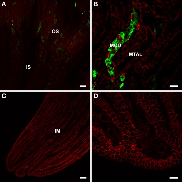
Figure 2.

BGT1 localization in the kidney. The kidney sections from wildtype and knockout were labeled with anti-BGT1 antibody (red; Ab#590; 1 μg/ml), and with fluorescein-conjugated D. biflorus agglutinin (green; 1:300; marker for collecting ducts). (A,B) are from the outer strip of outer medulla, and (C,D) are from the tip of the renal papilla. The images from the sections from knockout mice are not shown here. Scale bars for (A,C) = 50 μm; scale bars for (B,D) = 20 μm. Immunochemistry was performed using the same materials and procedures described in detail by Zhou et al. (2012a).
