
Figure 2.
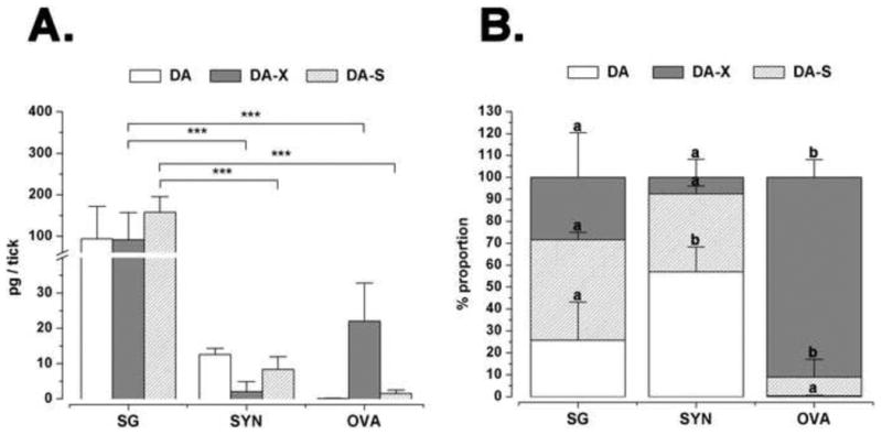
(A) Tissue-specific quantification of dopamine and its conjugates in the D5 tick salivary glands (SG), synganglia (SYN) and ovaries (OVA). Procedures for measuring the quantities of free dopamine (DA), conjugated dopamine (DA-X), and dopamine sulphate (DA-S) are in the text. (B) The data represent the proportions of dopamine and its conjugates in each tick organ. Values in the chart represent means of three biological replicates with error bars representing standard deviation (S.D.). In A and B, asterisks and letters indicate significant differences of p<0.001 and p<0.05, respectively, based on one-way ANOVA with Tukey post-hoc comparison.