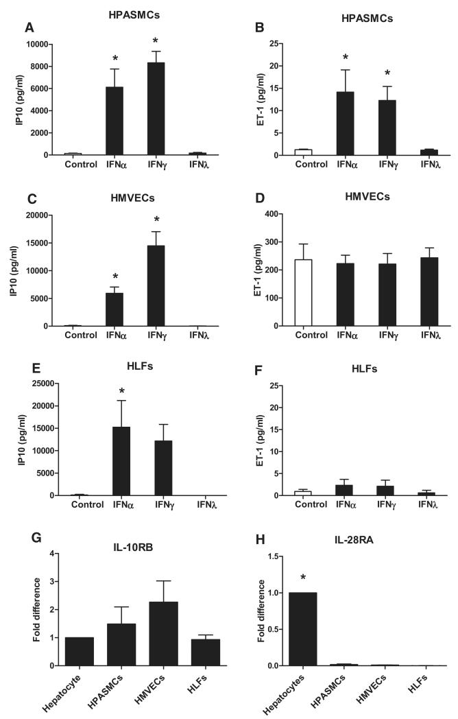
Figure 1.
Response of pulmonary vascular cells to types I, II, and III interferons (IFNs) and type III IFN receptor expression in pulmonary vascular cells as compared with hepatocytes. Human pulmonary artery smooth muscle cells (HPASMCs; A and B), human lung microvascular endothelial cells (HMVECs; C and D), and human lung fibroblasts (HLFs; E and F) were treated with IFNα (10 ng/mL), IFNγ (10 ng/mL), and IFNλ (1000 ng/mL) in the presence of tumor necrosis factor (TNFα; 10 ng/mL) and assayed for IFN γ inducible protein 10 (IP10; A, C, and E) and endothelin 1 (ET-1; B, D, and F). Data are presented as mean±SEM from n=3 to 6 experiments performed in singlicate. Statistical significance (*P<0.05) compared with control was determined by 1-way ANOVA with Dunnett multiple comparison post-test adjustment. IL10RB (G) and IL28RA (H) gene expressed as mean±SEM fold difference compared with hepatocytes from n=3 experiments in the absence of TNFα. Statistical significance (*P<0.05) compared with hepatocytes was determined by 1-way ANOVA with Dunnett multiple comparison post-test adjustment.

