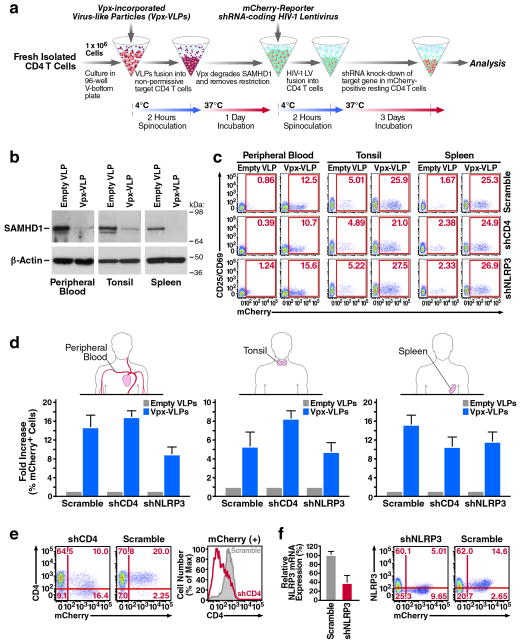
Figure 1.
Genetic manipulation of resting CD4 T cells by lentiviral vectors using sequential spinoculation of Vpx-VLP and shRNA-encoding HIV LV particles. (a) The spinoculation procedure. Fresh target cells were spinoculated with Vpx-VLPs, followed by shRNA-encoding HIV LV particles. Particles were pseudotyped with the CXCR4-tropic Env of HIV-1, which supports efficient fusion of virions to resting CD4 T cells. A detailed description is available in the Methods section (b) Spinoculation of Vpx-VLPs degrades SAMHD1 in resting CD4 T cells isolated from peripheral blood, tonsil, or spleen after 24 hours. (c) Efficient infection of resting CD4 T cells by three independent HIV LV particles indicated by mCherry transgene expression. Cells expressing mCherry remained viable (Supplementary Fig. 2) and negative for CD25/CD69 activation markers as indicated. (d) Fold increase of HIV LV infection after spinoculation with Vpx-VLPs as observed in CD4 T cells from peripheral blood, tonsil and spleen. (e) A marked decrease of surface CD4 expression in blood CD4 T cells productively infected with shCD4 HIV LV particles. (f) Reduced intracellular NLRP3 mRNA and protein expression in mCherry-positive CD4 T cells after infection with shNLRP3 HIV LV particles. Note that CD4 and NLRP3 expression were not reduced in mCherry-negative CD4 T cell populations. These data are representative of four independent experiments performed using CD4 T cells from peripheral blood, tonsil and spleen from four different donors.

