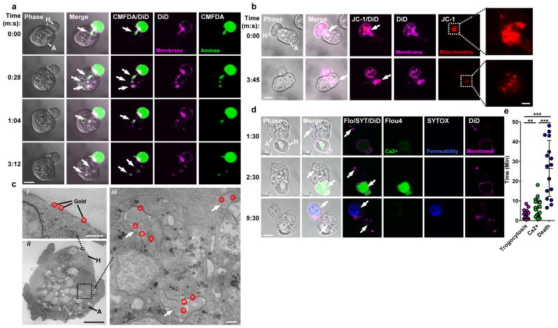
Figure 1. Amoebae internalize human cell bites, preceding human cell death.
a, DiD and CMFDA-labeled human Jurkat cells (H); an amoeba (A) internalizes bites (arrows) over time. Images are representative of three independent experiments. b, DiD and JC-1-labeled human Jurkat cells; mitochondria (arrow) are ingested by the amoeba in a bite. Images are representative of six independent experiments. c, i–ii, Electron microscopy with gold-labeled human Jurkat cells (gold, circles). iii, Bites (arrows) within an amoeba. Images are representative of two independent experiments. d, DiD and Flou4-labeled human Jurkat cells; with SYTOX blue present during imaging. Arrows, amoebic trogocytosis (1:30), [Ca2+]i elevation (2:30) and membrane permeabilization (9:30). Images are representative of 15 independent experiments. e, Timing in 60 cells from 15 independent experiments; shown are data points, means and standard deviations; P-values from t-tests: * < .05, ** < .01, *** < .001. Bars: 10 μm (a, b, d), 2 μm (b, insets), 0.5 μm (c, i, iii) and 5 μm (c, ii).

