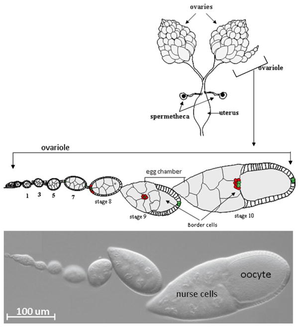
Fig. 1.
Anatomy of the Drosophila ovary. Top – Schematic drawing of a pair of ovaries dissected from female fruit fly. A schematic drawing of an enlarged single ovariole containing egg chambers of the indicate stages of development. The bottom panel shows a DIC image of an ovariole with similar stages of egg chamber development.

