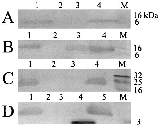
FIG. 2.
Identification of Gvp proteins by immunoblot analysis. (A) Whole-cell lysates of Halobacterium sp. strain NRC-1 (lane 1), SD109 (lane 2), and SD109 (pFL2gvpJ::κ1) (lane 3) and purified gas vesicles (lane 4) were electrophoresed, transferred to membrane, and probed with GvpJ antibodies. (B) As in panel A, except lane 3 contains whole-cell lysate of Halobacterium sp. strain SD109(pFL2gvpM::κ1) and the blot was probed with GvpM antibodies. (C) As in panel A, except lane 3 contains whole-cell lysate of Halobacterium sp. stran SD109(pFL2gvpF::κ5) and the blot was probed with GvpF antibodies. (D) As in panel A, except lane 3 contains whole-cell lysate of Halobacterium sp. strain SD109(pFL2gvpG::κ1), lane 4 contains synthetic GvpG peptide, and the blot was probed with GvpG antibodies. Prestained protein standards are displayed in lanes marked M, and molecular masses are indicated.

