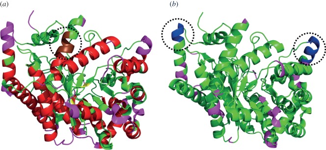
Figure 5.

Prediction of local conformational rigidity and flexibility. Structure of glutamate mutase [PDB ID: 1CCW, chain B] [218] (a) highlighting the predicted secondary structures [219]: helices (red) and strands (yellow), flexible regions [217] (pink) and a discordant helix with high strand propensity (brown, highlighted within the dotted circle) [215]. (b) The flexible regions predicted (probability > 0.5 and confidence > 10) by PredyFlexy method [216] (pink) and the predicted disordered region [220] is shown in blue (highlighted within the dotted circles). (Online version in colour.)
