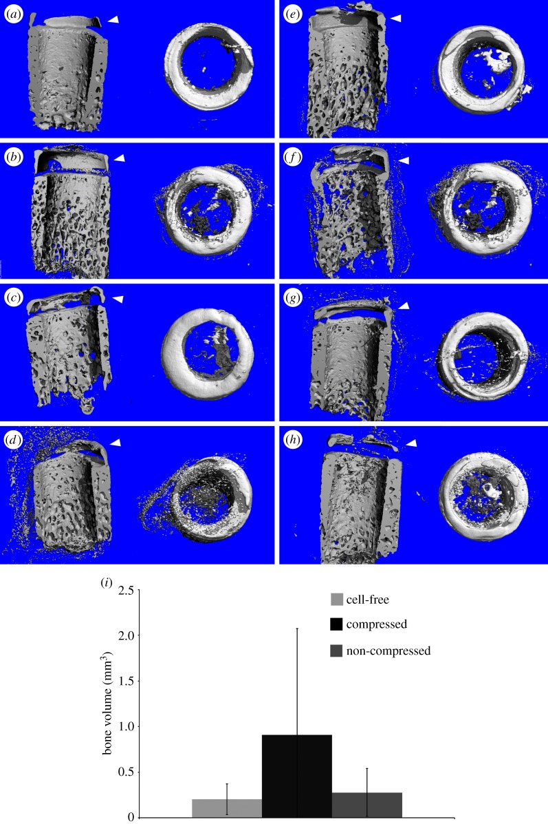
Figure 4.
µCT images showing mineralized tissues in the scaffold. Explants of the triphasic scaffold inserted in bovine core, which had cell-free (osteoblast) (a,e) and osteoblast-seeded bone compartments (patient 1: b,f; patient 2: c,g; patient 3: d,h), had little bone formation in the osseous compartment. Explants that contained alginate gels subjected to a one-week compressive loading prior implantation are shown in (e–h), and those that had non-loaded alginates are shown in (a–d). Mineralization was visible on the outer rims of the cartilage in all groups including the cell-free controls. (i) Bone volume within the bovine osteochondral defect as measured by µCT. There was no statistical difference among the three groups (cell-free n = 4, Comp and NC n = 14). (Online version in colour.)

