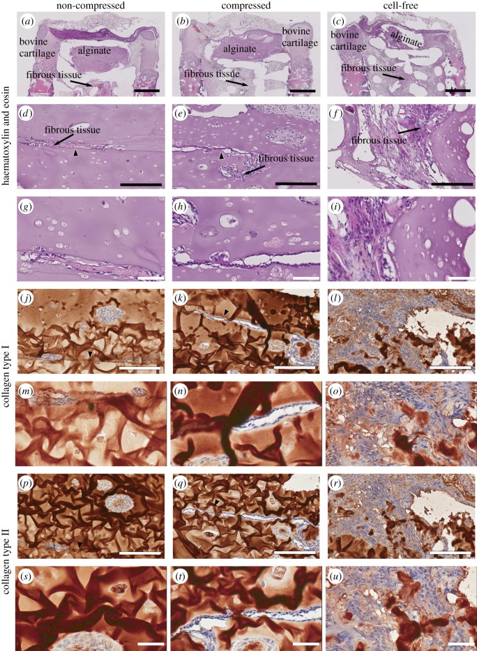
Figure 6.
Histological and immunohistochemical images of explant cartilaginous compartment. While explants implanted with both NC (a,d,g,j,m,p,s) and Comp (b,e,h,k,n,q,t) alginate gels had retained much of the alginate constructs during the 12-week in vivo culture, those in the cell-free group had little alginate left (c,f,i,l,o,r,u). While S and MD alginate gels remained intact in the cell-seeded groups, fibrous tissue infiltration was visible in the interface between the two gels (arrows). Cells found within the alginate compartment in all groups formed dense aggregates but did not stain strongly for either collagen type I (j–o) or II (p–u). Scale bars: (a–c), 1 mm; (d–f, j–l, p–r), 200 µm; (g–j, m–o, s–u), 50 µm. (Online version in colour.)

