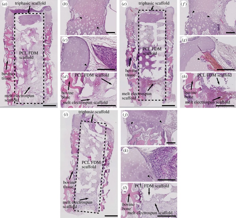
Figure 8.
Haematoxylin-/eosin-stained sections of osteochondral explants. (a–d) NC, (e–h) Comp and (i–l) cell-free. Fibrous tissue infiltration was seen in both the inner and outer edges near the lower portion of the bovine cartilage in all explants (a,e,i, and arrowheads in b,f,j). Fibrous tissue infiltration was also seen in the upper part of the cartilage and they were seen primarily from the inner edges of the cartilage (arrowheads in c,g,k). Fibrous tissues also fully infiltrated the bovine bone, PCL-FDM scaffolds and the PCL mesh (a,e,i,d,h,l). Scale bars: (a,e,i), 1 mm; (b–d,f–h,j–l), 200 µm. (Online version in colour.)

