Figure 1.

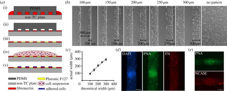
Experimental model. (a) Schematic of cell patterning procedure; (i) PDMS moulds are placed onto a non-tissue culture-treated plate, and an FN solution fills the channels of the mould. (ii) After 1 h of incubation, the FN solution is aspirated. (iii) After drying, the PDMS mould is removed and the plate is treated with Pluronic, which coats the non-patterned surface and leaves it non-adherent. (iv) A drop of cell suspension is spread over the patterned area. (v) The cells adhere to the FN patterns only. (b) Phase contrast micrographs of pre-chondrogenic cells cultured on linear adhesive islands or non-patterned substrates taken after 24 h in culture. The island width varies from 100 to 300 μm, while the non-adhesive space between islands is 300 μm wide for all conditions. (c) Plot indicating the correlation between the theoretical island width and the actual island width measured during cell culture. (d) Fluorescent micrographs of pre-chondrogenic cells isolated from chicken limb buds cultured for 2.5 days on 150 μm wide adhesive islands, showing a representative mesenchymal condensation. Cell nuclei are stained blue (DAPI), green indicates positive staining with PNA lectin, and cell-deposited fibronectin is stained red. Scale bar, 100 μm. (e) Fluorescent micrographs of pre-chondrogenic cells cultured for 2.5 days on 100 μm wide adhesive islands, showing a representative mesenchymal condensation stained with PNA lectin (green), and for NCAM (red). Scale bar, 50 μm. (Online version in colour.)
