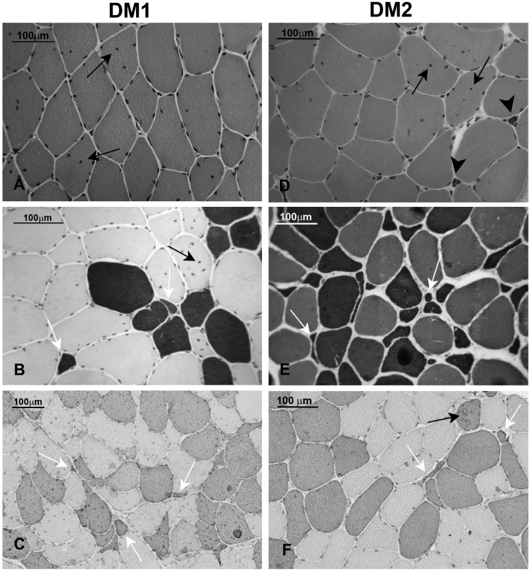
Figure 2.
Panel showing muscle histology in DM1 and DM2. A-C. Transversal sections from DM1 muscle biopsies. A. Haematoxylin & Eosin: fiber size variation and central nuclei (arrows) are present. B, C. The population of atrophic fibers (white arrow) are preferentially type 1 fibers as demonstrated in sections stained for ATPase pH 4.3 (B, dark brown) or immunostained for myosin MHCslow (C, brown). Black arrow indicate centrally located nuclei. D-E Transversal sections from DM2 muscle biopsies. D. Haematoxylin & Eosin: as in DM1 muscle, fiber size variation and central nuclei (arrows) are present. Abundant nuclear clumps are also present (arrow heads) despite the muscle shows an early stage pathology. E, F. Type 2 fibers are predominantly affected in DM2 muscle: in routine laboratory muscle staining such as ATPase pH 10.0 (E) or immunostaining for myosin MHCfast (F), type 2 fiber atrophy (white arrows) and type 2 central nucleation (black arrow) are commonly observed.

