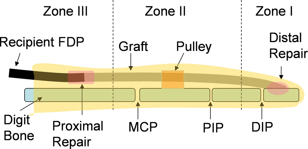
Figure 1.
Schematic diagram of the graft with digits zones I-III. The graft in zone I and zone II was assessed for work of flexion and adhesion formation. The graft in zone I was also tested for tendon to bone healing strength, and the graft in zone II was assessed for graft friction. The proximal host tendon to graft repair in zone III was used to evaluate the adhesion breaking strength. The graft segments in zone I, II, and III were also processed for histology. (DIP: distal interphalangeal joint; PIP: proximal interphalangeal joint; MCP: metacarpophalangeal joint)

