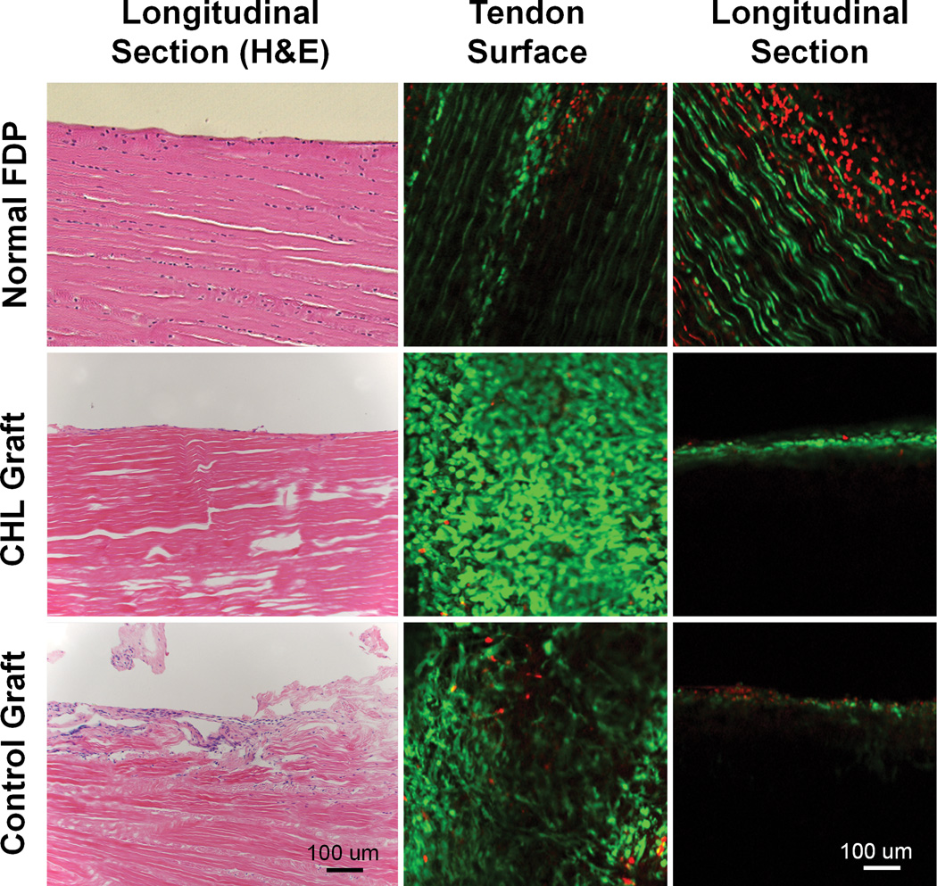
Figure 7.
Left column displays H&E staining of normal FDP tendon (top, left) Note that tenocytes are aligned along tendon fascicles, with a smooth surface. The graft treated with CHL also showed a smooth surface with a cell layer attached (Center, left). No cells are present within the CHL grafts. The saline treated graft show a rough surface with adhesion formation (bottom, left).
Middle column (tendon surface) and right column (tendon longitudinal section) show viable cells (green) identified by calcein-AM and ethidium homodimer probe under confocal microscopy. All three groups showed a large amount of live cells on the tendon surface. Cells are well aligned longitudinally in the normal FDP tendon (top, middle). However, the cells on both grafts are disorganized (center, middle and bottom, middle). In the tendon longitudinal sections, there are cell layers within the normal FDP tendon (top, right). Many dead cells (in red) were also observed on the normal tendon longitudinal section, which might be related to sample preparation (sectioning or manipulating) before staining that caused the cell death. No viable cells or dead cells were observed within either graft tendons (center, right and bottom, right).

