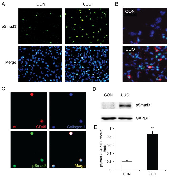
Figure 1. Smad3 is activated in myeloid fibroblasts in the kidney after obstructive injury.
A. Representative photomicrographs of kidney sections stained for phospho-Smad3 (green) and counter stained with DAPI (blue) (Original magnification: X400). B. Representative photomicrographs of kidney sections stained for phospho-Smad3 (green) and DDR2 (red) and counter stained with DAPI (blue) (Original magnification: X600). C. Representative photomicrographs of isolated renal fibroblasts stained for CD45 (red), phospho-Smad3 (green), collagen I (blue). D. Representative Western blots show activation of Smad3 in UUO kidneys of WT mice. E. Quantitative analysis of Smad3 phosphorylation in the control and UUO kidneys of WT mice. ** P < 0.01 vs WT controls. n=6.

