Figure 6.
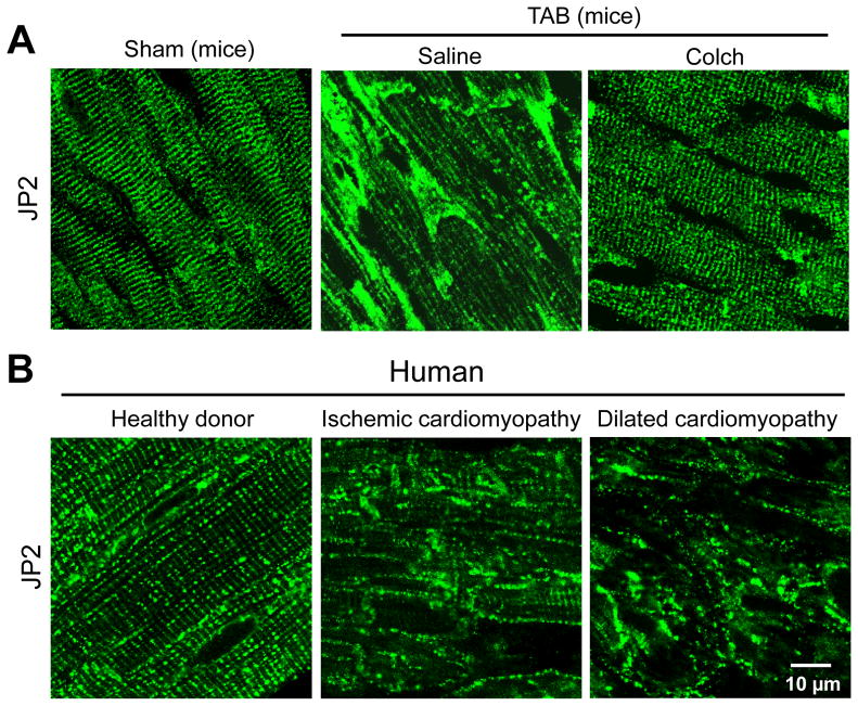
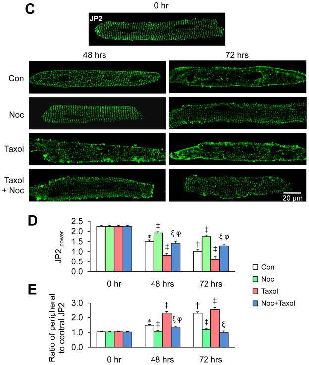
Microtubule polymerization accelerates JP2 redistribution in response to stress. A. Representative confocal images of in situ JP2 immunofluorescent staining in LV cryosections from age-matched sham and TAB mice injected with saline or colchicines (Colch). (n=3–4 hearts per group). B. Representative in situ JP2 immunofluorescence in LV cryosections from human “healthy” donor hearts (n=3), and hearts with ischemic (n=4) or dilated cardiomyopathy (n=3). 10–15 confocal images were acquired from each heart sample. C. Representative images of isolated cardiomyocytes stained with anti-JP2 antibody after treatment with 10 μM nocodazole, 20 μM taxol, or the combination of both in culture for 48 or 72 hrs. D. Average JP2power in cultured cardiomyocytes with different treatments. E. Semi-quantitative analysis of JP2 redistribution from T-tubule membrane to periphery sarcolemma. Con: DMSO control; Noc: nocodazole. n = 15–20 cells per group.* p<0.05 vs control at 0 hr; † p<0.05 vs control at 48 hrs; ‡ p<0.05 vs control at the same time point; ξ p<0.05 vs Taxol of the same time point; φ p<0.05 vs Noc at the same time point. p<0.001 among Con, Noc, Taxol, and Noc+Taxol groups at 48 hrs and 72 hrs (by Linear mixed-effects model).


