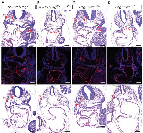
Figure 1. Heg and Ccm2 interact within endothelial cells during embryonic development.
H&E staining and Immunostaining with anti-Pecam antibody of transverse sections of E9.5 embryos reveal the presence of normally lumenized dorsal aortas (DA), cardinal veins (CV) and branchial arch arteries (BAA) in the Tie2Cre+;Heg1fl/−(A) and Heg−/−;Ccm2fl/+(C) embryos but not in Tie2Cre+;Heg1fl/−Ccm2fl/+(B) or Heg−/−;Ccm2+/−(D) embryos. H&E staining of transverse sections of caudal region of E9.5 embryos also revealed dilated cardinal veins and non-lumenized dorsal aortas in Tie2Cre+;Heg1fl/−Ccm2fl/+(B), and Heg−/−;Ccm2+/−(D) embryos. AO Sac, aortic sac; CV, cardinal vein; DA, dorsal aorta; BAA, Branchial arch ateries. Scale bars, 100μm.

