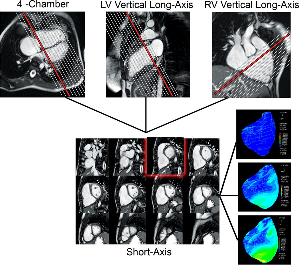
Figure 1.
CMR assessment of biventricular volumes and mass in a patient with repaired TOF. Cross-referencing between ventricular long- and short-axis imaging planes aids determining inclusion of basal slices in the ventricular volume analysis. Right lower panel: 3D strain maps of the RV at end-diastole (top), mid-systole (middle), and late systole (bottom).

