Figure 7.
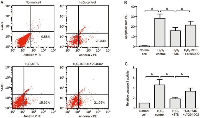
The H2O2-induced in vitro effect of STS on the apoptosis and caspase 3 activity of neonatal rat cardiac cells. (A) STS (3 μg/mL) was pretreated at 12 h before the incubation of cells with 200 μmol/L of H2O2 for 1 h. Normal cells; control cells exposed to 200 μmol/L of H2O2 for 1 h; 3 μg/mL STS-pretreated cells at 1 h after exposure to 200 μmol/L of H2O2 (c); 3 μg/mL STS and 10 μmol/L LY294002 co-pretreated cells at 1 h after exposure to 200 μmol/L of H2O2 (d). (B) Statistical analysis of the flow cytometric analysis data. (C) Statistical analysis of relative caspase 3 activity. The data represent the mean±SD. bP<0.05 was considered statistically significant among each group.

