Figure 3.
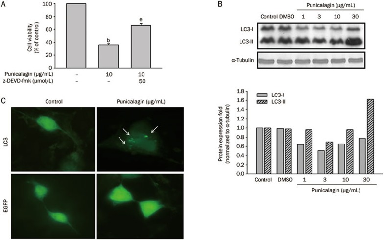
Punicalagin induces autophagic cell death in U87MG cells. (A) Cells were pretreated with or without z-DEVD-fmk for 15 min prior to treatment with 10 μg/mL punicalagin for 24 h. Cell viability was determined using an MTT assay, and the viability of the untreated cells was measured as the 100% control. The data are presented as the mean±SEM of triplicate experiments. bP<0.05 vs the 100% control. eP<0.05 vs the 100% control and the inhibitor control. (B) Cells were treated with 10 μg/mL punicalagin for 24 h, and the lysates were collected and immunoblotted with antibodies specific for LC3 and α-tubulin. Protein quantitation data are located in the right panel and were normalized to α-tubulin. (C) Cells were transfected with GFP-LC3 or enhanced green fluorescence protein (EGFP) and were then treated with 10 μg/mL punicalagin for 24 h. All images were taken with an Olympic fluorescence microscope (Model IX71) immediately after fixation by paraformaldehyde. Arrows indicate the punctate dots.

