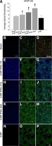
Figure 4.
Optical densitometry of GFAP from the transplant site. Astrocytes (GFAP; green) were observed around the transplanted iPSCs (blue), but no colocalization was observed between the two labels. A significant between-group difference in the average optical densitometry of GFAP (A) was observed. It was revealed that 3-NP rats receiving iPSC transplant at either 21 or 42 days displayed significantly higher GFAP labeling than sham rats and 3-NP rats that received transplantation of iPSCs at 7 days. The 3-NP rats receiving iPSC transplantation at 7 days displayed significantly more astrocyte activation than 3-NP rats that did not receive transplants. Sham rats are shown in (B–D); 7-day iPSC rats are shown in (E–G); 21-day iPSC rats are shown in (H–J); 42-day rats are shown in (K–M); 3-NP rats that did not receive transplants are shown in (N–P). (Scale bar = 50 μm; bar graph represents mean value; error bars represent SEM; ∗, significantly different from sham rats, p < .05; †, significantly different from 3-NP rats, p < .05; #, significantly different from 3-NP iPSC 7-day rats, p < .05.) Abbreviations: 3-NP, 3-nitropropionic acid; GFAP, glial fibrillary acidic protein; iPSC, induced pluripotent stem cell; OD, optical density.

