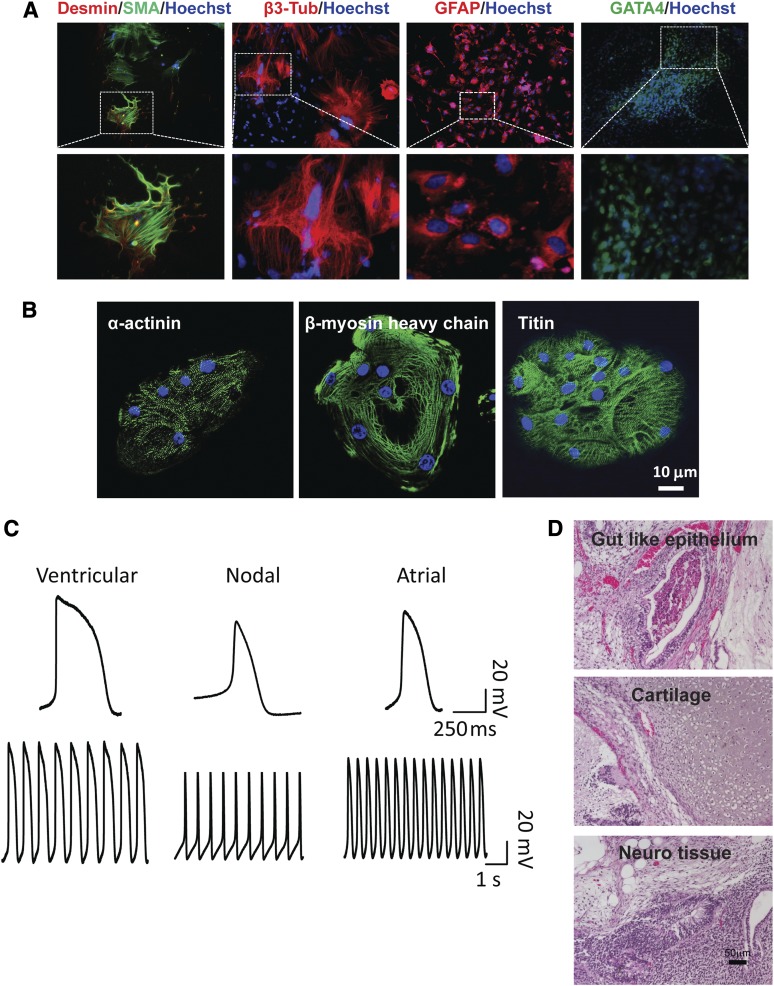
Figure 2.
Finger-prick-derived induced pluripotent stem cells (FPiPSCs) differentiate into derivatives of three germ layers. (A): Immunofluorescent staining of differentiation markers after in vitro differentiation of FPiPSCs. Ectoderm was stained by GFAP and β3-tubulin antibodies, whereas endoderm was stained by antibody against GATA4. All images in the upper panel were acquired with original magnification ×10 objectives, except for GATA4 staining image, which was acquired with ×4 objectives. Lower panel images are shown in higher magnification. Hoechst staining indicates the nucleus. (B): Immunofluorescent staining of α-actinin, β-myosin heavy chain, and titin in human induced pluripotent stem cell (hiPSC)-derived cardiomyocytes. Hoechst staining indicates the nucleus. (C): Patch-clamp analyses of hiPSC-derived cardiomyocytes. (D): Hematoxylin and eosin staining of teratomas derived from immune-deficient mice injected with FPiPSCs shows tissues representing all three embryonic germ layers, including gut-like epithelium (endoderm), cartilage (mesoderm), and neural tissue (ectoderm). Scale bar = 50 μm. Abbreviations: β3-Tub, β3-tubulin; GFAP, glial fibrillary acidic protein; SMA, smooth muscle α actin.

