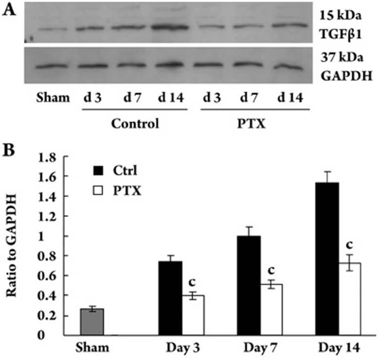
Figure 3.
Expression of TGFβ1 protein in kidney tissue. (A) Representative western blot of TGFβ1 in sham and UUO rat treated with or without PTX. (B) Compared with control group, PTX treatment inhibited the increased expression of TGFβ1. The data was shown as ratio of TGFβ1 density to GAPDH density. The values were expressed as mean±SD of 3 independent experiments. cP<0.01 vs control.

