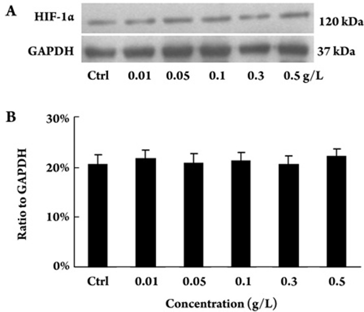
Figure 7.
Effect of PTX on expression of HIF-1α in cultured HK-2 tubular epithelial cells. HK-2 cells were treated with indicated concentration of PTX for 72 h. The expression of HIF-1α was determined with Western blot. (A) Representative Western blot of HIF-1α in HK-2 cells. (B) Compared with control, PTX treatment did not change the expression of HIF-1α. The data were shown as ratio of HIF-1α density to that of GAPDH and expressed as mean±SD of 3 independent experiments. P=0.935 vs control by ANOVA.

