Fig. 3.
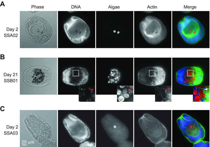
Localization of algae in Aiptasia larvae. Phase-contrast (Phase) and deconvolved fluorescence images (see Materials and methods) of larvae exposed to compatible (A,B) or incompatible (C) algal types as in Fig. 2. DNA, Hoechst staining of nuclei (blue in Merge); Algae, chlorophyll autofluorescence of algal cells (red in Merge); Actin, staining of host cell cytoplasmic actin with AlexaFluor 488-conjugated phalloidin (green in Merge). (A,B) Accumulation of compatible algae within the gastrodermal cells of the host. Inset, a region in which a host nucleus (arrowheads) and two algal cells (or one dividing cell) can be seen surrounded by phalloidin-stained host cytoplasm. (C) Presence of incompatible algae within the gastric cavity but not the gastrodermal cells. Scale bar applies to all panels.

