Figure 1.

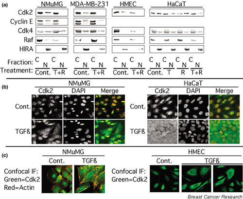
TGF-β alters Cdk2 localization. (a) NMuMG, MDA-MB-231, primary human mammary epithelial (HMEC), or HaCaT cells were treated for 24 h with normal growth medium (Cont.), 10 ng/ml TGF-β1 (T), 100 nM rapamycin (R), or a combination of both (T + R). The cells were subjected to subcellular fractionation into nuclear (N) and cytoplasmic (C) extracts as described in Materials and Methods and the resulting fractions were analyzed by immunoblotting with the indicated antibodies. HIRA served as a marker for the nucleus, and Raf was used as a marker for the cytoplasm. (b) NMuMG cells (left panel) or HaCaT cells (right panel) were treated for 24 h with 10 ng/ml TGF-β1 and the cells were stained with antibodies specific for Cdk2 (shown in green), and with 4',6'-diamidino-2-phenylindole (DAPI; shown in red) as a nuclear stain, as described in Materials and Methods. In the merged images, Cdk2 and DAPI staining are overlaid. (c) NMuMG cells (left panel) treated as in (b) were stained for Cdk2 (green) and actin (red) and were imaged by confocal microscopy. HMECs (right panel) treated as in (b) were stained for Cdk2 (green) and were imaged by confocal microscopy.
