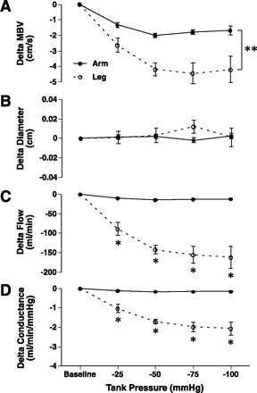
Fig. 3.

Absolute MBV, diameter, flow, and conductance changes (Δ) under sustained increases in tank suction (−25, −50, −75, and −100 mmHg) in the brachial and femoral arteries. Legs compared with arms had greater velocity reductions in response to changes in suction (A) with no change in diameters in either limb (B). Leg flow and conductance changes were greater at every tank suction level compared with the arm (C and D). Baseline, baseline levels at ambient pressure where Δ = 0; • and solid line, arm; ○ and dashed line, leg; *Significant difference between arm and leg at a specific negative tank pressure. **Significant main effect difference between arm and leg.
