Figure 2.

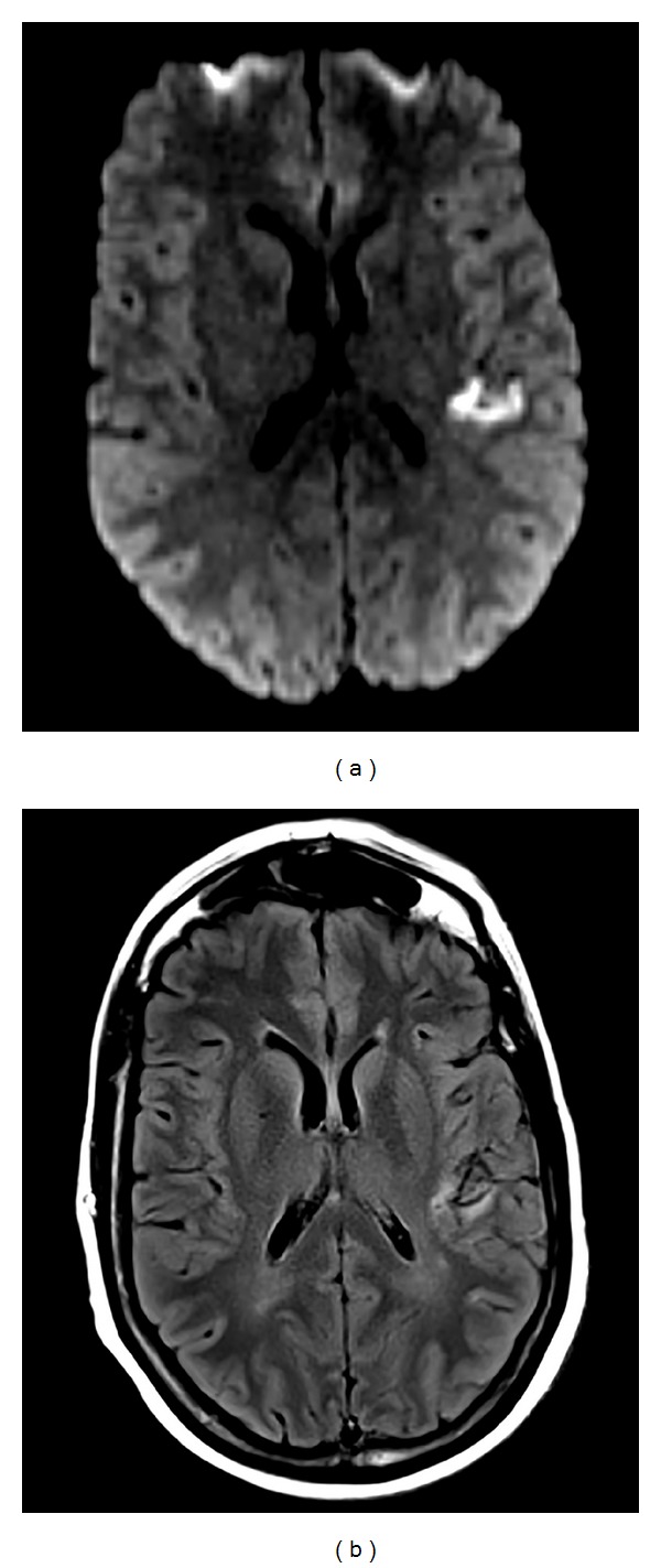
Magnetic resonance imaging (MRI) shows abnormal reduced diffusivity (a) and T2 hyperintensity (b) in the left posterior insula and temporoparietal operculum.

Magnetic resonance imaging (MRI) shows abnormal reduced diffusivity (a) and T2 hyperintensity (b) in the left posterior insula and temporoparietal operculum.