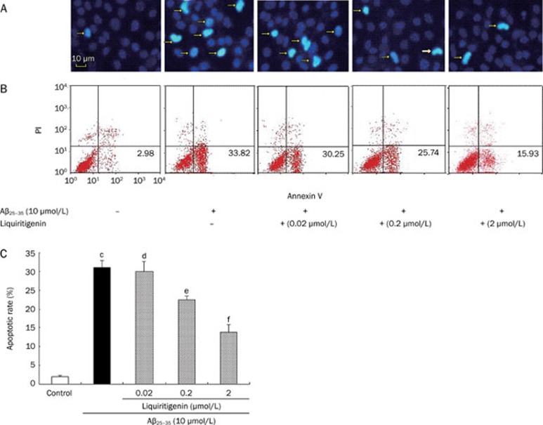
Figure 3.
Liquiritigenin protects neuron cells against Aβ25–35-induced apoptosis. (A) Nuclei were stained with fluorescent dye Hoechst 33342 for assessment of apoptosis; arrowheads indicate apoptotic cells. Magnification: ×200. (B) Representative FCM histograms stained by Annexin V-FITC/PI. Annexin V+ and PI− cells were designed as apoptotic. (C) Apoptotic rate of cells, determined by FCM. Bars represent mean±SEM (n=4). aP>0.05, bP<0.05, cP<0.01 vs control; dP>0.05, eP<0.05, fP<0.01 vs Aβ25–35 alone.

