
Fig. 2. N-nitrosation in the XO/HX/SPENO system.
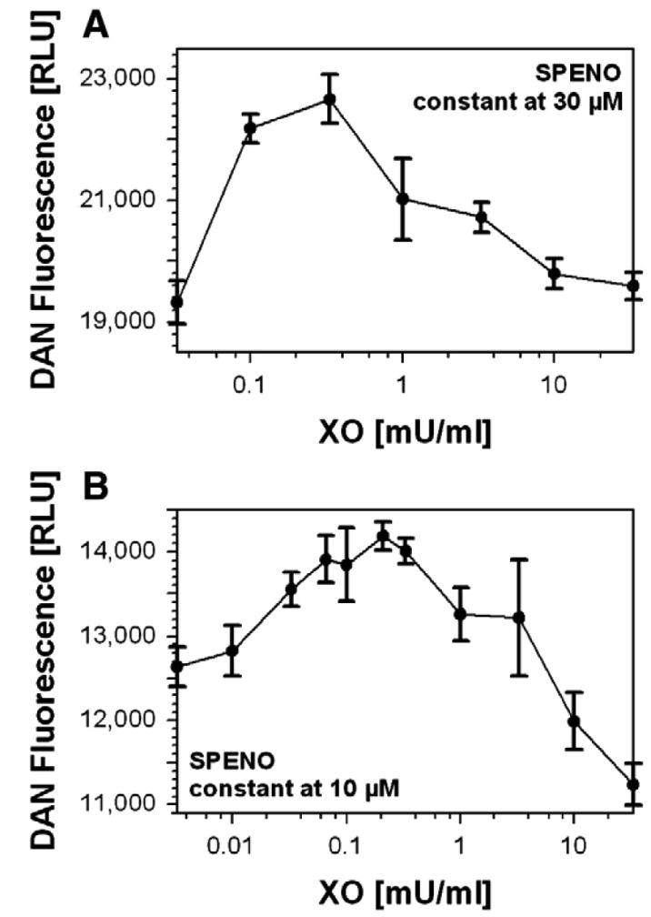
(A) DAN (100 μM) derived fluorescence was monitored at constant SPENO (30 μM) and increasing XO concentrations in 0.1 M potassium phosphate buffer, pH 7.4, containing 1 mM HX at 37°C. Data were collected after 5 min incubation. (B) DAN (20 μM) fluorescence was monitored at constant SPENO (10 μM) and increasing XO concentrations in 0.1 M potassium phosphate buffer, pH 7.4, containing 1 mM hypoxanthine at 37°C. Data were collected after 25 min incubation and are expressed as the means±SEM of two or three independent experiments.