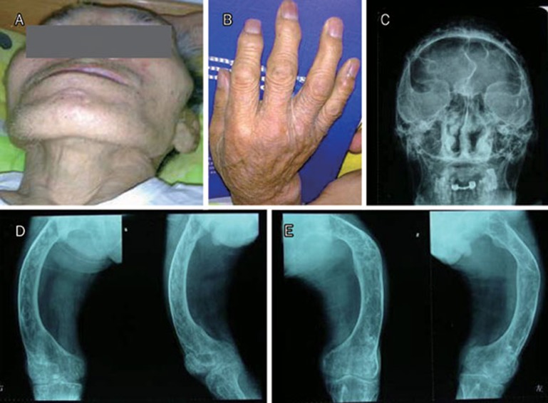
Figure 3.
(A) Photograph of patient II1 showing facial deformity with maxillary expansion. (B) Photograph of the hand of the same patient showing swelling of the proximal interphalangeal and distal interphalangeal joints. (C) Skull radiograph showing osteolysis and osteosclerosis. (D) and (E) Right and left femora radiographs showing deformity and bony expansion with osteolysis and osteosclerosis.

