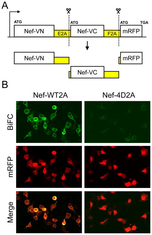
Figure 1.
Single-plasmid expression vector for detection of Nef-BiFC inhibitors by HCS. (A) The coding regions for the two fusion proteins constituting the Nef BiFC pair (Nef-VN and Nef-VC) as well as the mRFP reporter protein are transcribed as a single mRNA separated by the viral ribosomal skipping sequences, E2A and F2A. The 2A sequences cause discontinuous translation, resulting in the three distinct proteins illustrated. (B) Validation of the single-plasmid biosensor (Panel A) for Nef dimerization by BiFC. Human 293T cells were transfected with single-plasmid BiFC vectors for detection of wild-type Nef dimerization (Nef-WT2A) as well as dimerization-defective Nef (Nef-4D2A) as a negative control. Forty-eight hours later, the cells were imaged by confocal microscopy to detect Nef dimerization as reconstituted YFP fluorescence (BiFC) as well as expression of the mRFP reporter. A merged image is also shown.

