
Figure 2. Transgenic kittens.
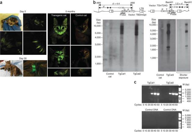
(a) Ambient light– and 485 nM light–illuminated images showing GFP signal at indicated times after birth for TgCat3. In the 30 d and 5 month images TgCat3 was photographed with a non-transgenic control cat (right). Coat, claw, whisker, nose, tongue and oropharyngeal mucosa fluorescence are evident; fluorescence was relatively quenched in dark fur. (b) Southern blotting of genomic DNA from TgCat1, TgCat2 and TgCat3. Southern junction blot designs are shown. d, distance from vector-host DNA junction to nearest genomic AflIII or BamH1 site in base pairs; P, promoter; LTR, long terminal repeat; G, eGFP; T, TRIMCyp. Genomic DNA from tail tips was digested with AflIII (left blot). Genomic DNA from peripheral blood mononuclear cells was digested with BamH1 (right blot). After electrophoresis and Southern blot transfer, membranes were probed for integrated vector DNA as indicated. (c) Amplicons from semiquantitative PCR amplifications of kitten genomic DNA using primers for the rhTRIMCyp sequence. M, marker. Cycles, number of PCR amplification cycles. Quantitative PCR showed that TgCat1 and TgCat2 had 15.2 ± 2.1 and 4.38 ± 0.2 GFP gene copies per cell equivalent respectively, using a value of 6.3 pg genomic DNA per diploid cell and normalizing to the signal obtained with GAPDH primers.