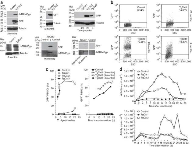
Figure 3. Immunoblotting and FIV challenge of transgenic PBMCs.

(a) Representative immunoblots for GFP and HA-tagged rhTRIMCyp in PBMCs isolated from transgenic and control cats. All PBMC are activated (PHA-E) except for the TgCat1 sample labeled 'unactivated'. (b) Flow cytometry analysis of GFP expression in activated PBMCs. Percentages of cells that are GFP-positive are indicated. (c) GFP expression in PBMCs versus cat age (left) and GFP expression in PBMCs from a single time point, as a function of days in ex vivo culture; sampling here was at 3–4 months of age (arrow). (d) PBMCs from cat were infected with 105 Crandell feline kidney cells (CrFK) cell-infectious units of FIV on day 0, washed on day 1 and then followed by sampling for supernatant reverse transcriptase activity determination every 48 h as shown. RT, reverse transcriptase; SSC, side scatter.
