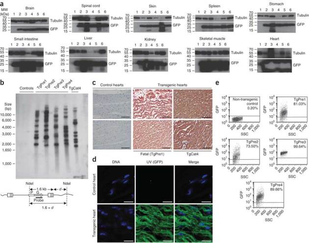
Figure 5. Whole body analyses of TgCat4 and late developmental stage fetuses.

(a) Immunoblotting on lysates from indicated organs from non-transgenic control cat (lanes 1); preterm fetal tissues (lanes 2–5; and TgCat4 (lanes 6). Uncropped versions of these films are available in Supplementary Figure 4. These are minimal (<1 s) film exposures of the immunoblots; the central white-out in the heart GFP band is a result of heavy GFP expression causing artifactual exhaustion of chemiluminescent substrate. (b) Southern blotting for integrated vector DNA. Genomic DNA from heart tissue was digested to completion with NdeI and 5 μg were loaded per lane. Specific bands for intact integrated vector are predicted to be ≥ 1.6 kb. Feline T cell line (FetJ) (left control); control cat; TgPre1–4 from pregnancy C; and TgCat4 are shown. (c) Cardiac muscle from a control cat, TgPre1 and TgCat4 was subjected to indirect immunofluorescence with a monoclonal antibody to GFP. (d) GFP imaged directly in fresh thin sections of TgPre1 myocardium by epifluorescence microscopy. (e) FACS analyses of fetalPBMCs. Scale bars, 100 μm (black bars) and 20 μM (white bars).
