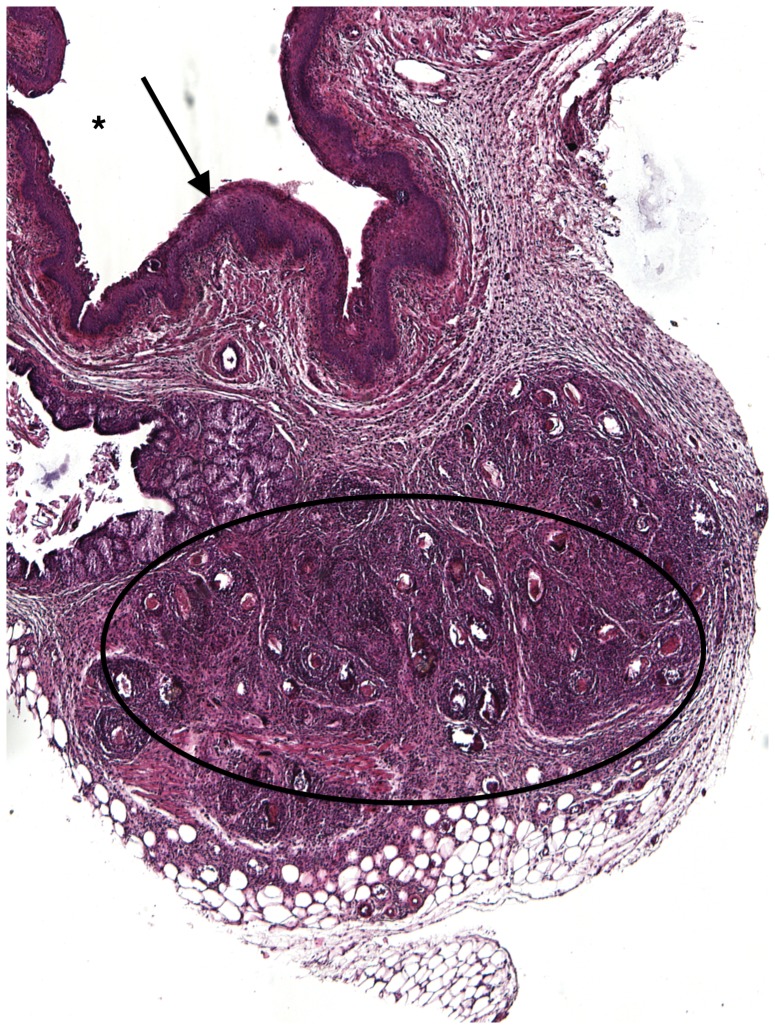
Figure 2. H&E Mouse vaginal walls injected with S. haematobium eggs post infection at 2 weeks.
Injections at all time points resulted in formation of schistosome egg-based granulomata, which consist mainly of eosinophils, neutrophils, plasma cells, lymphocytes, and epithelioid cells. Asterisk = Vaginal lumen, Arrow = vaginal epithelium, Circle = granuloma.

