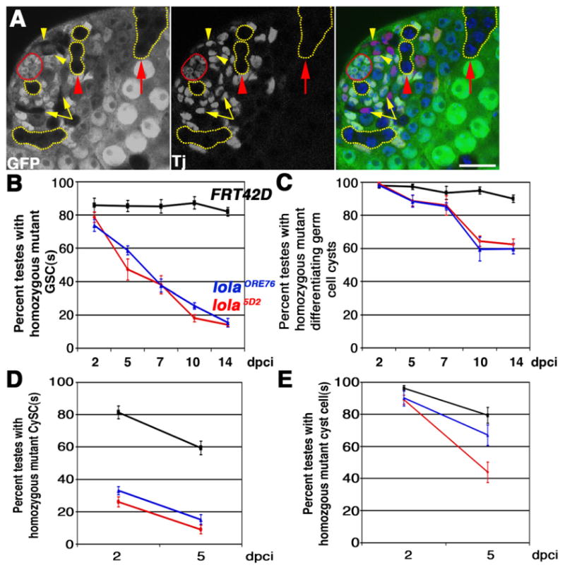
Fig. 2.

lola is required cell autonomously for GSC and CySC maintenance: (A) lolaORE76 mosaic testis containing clones marked by absence of GFP at 2 days post clone induction (dpci). Red: Tj, somatic nuclei. Green: Ubi-nls-GFP. Blue: DAPI. Red outline: hub. Yellow outlines: Examples of GFP negative germline clones. Note lola mutant GSC abutting the hub. Yellow arrowheads: lola mutant CySCs next to the hub. Yellow arrows: lola mutant cyst cells. Red arrowhead: lola mutant spermatogonial cyst. Red arrow: lola mutant spermatocyte cyst. Scale bar: 20 μm. (B) and (C) Percentage of mosaic testes containing homozygous mutant (B) GSC(s) or (C) spermatogonia and/or spermatocyte cyst(s) from 2 to 14 dpci. (D) and (E) Percentage of mosaic testes containing homozygous mutant (D) CySCs or (E) cyst cell(s) from 2 to 5 dpci. (B)–(E): Black: FRT42D control clones. Blue: lolaORE76 clones. Red: lola5D2 clones. Error bars: standard error of the mean. n≥75 testes per genotype, per time point.
